|
The INTERNATIONAL JOURNAL of APPLIED RESEARCH In Veterinary Medicine |
 |
| Current Issue |
| Previous Issues |
| Reprint Information |
| Back to The International Journal of Applied Research in Veterinary Medicine |
Comparative Studies on the Occurrence and Distribution of Epizootic Lymphangitis and Ulcerative Lymphangitis in Ethiopia
Bojia Endebu, DVM*
François Roger, MSc, DVM, PhD
*The Donkey Sanctuary Working Word Wide, The Donkey Health and Welfare Project, Faculty of Veterinary Medicine, Addis Ababa University, P. O. Box 34, Debre Zeit, Ethiopia
CIRAD-EMVT, Animal Health Programme TA 30/G, Campus
International de Baillarguet, 34398 Montpellier Cedex 5, France
KEY WORDS: equine, lymphangitis, Histoplasma farciminosum, Corynebacterium pseudotuberculosis, Ethiopia
Abstract
A study was conducted to determine the occurrence and distribution of epizootic lymphangitis (EL), caused by Histoplasma farciminosum, and ulcerative lymphangitis (UL), caused by Corynebacterium pseudotuberculosis, among equids (carthorses and mules) in different regions of Ethiopia. Data from 17 regional veterinary clinics located in different parts of the country were analyzed retrospectively. In addition, a monthly incidence study on these diseases in the 1100 carthorses in the towns of Debre Zeit and Akaki was conducted over 5 months. The cross-sectional study during the month of October indicated a prevalence of 5.0% (30 of 600) and 1.2% (5 of 100) in Debre Zeit and Akaki, respectively. Clinical, direct microscopic, bacteriologic, and mycologic examinations were used to confirm the cases. The retrospective studies indicated that the diseases occurred in large parts of the country, EL being more prevalent than UL. The percentage incidence and the period prevalence of EL in Debre Zeit were 2.2% and 10.1%, respectively, whereas in Akaki, they were 1.0% and 4.4%, respectively. The highest numbers of cases were observed in October (30 of 1000) and January (22 of 1000). The peak transmission season for EL coincided with the breeding season for flies. Because both diseases have significant economic and veterinary importance, further epidemiologic investigations are indicated to design optimal control strategies.
INTRODUCTION
The equine population in Ethiopia is estimated to be 2.75 million horses, 5.02 million donkeys, and 0.63 million mules. They are distributed in almost all the agro-ecologic zones of the country at an average density of 6.1 km-21 and account for 13.7% of the total biomass of domestic herbivores.2 Equids are mainly used as draught and pack animals. In some parts of Ethiopia, they are also used for ploughing.
There are a number of diseases that affect equids, thereby reducing the power they can provide. Epizootic lymphangitis (EL) and ulcerative lymphangitis (UL) are 2 important health problems that mainly affect horses. EL is a disease caused by the dimorphic fungus, Histoplasma farciminosum. It is characterized by suppurative inflammation of cutaneous lymphatic vessels, lymph nodes, and adjacent skin. UL is a contagious disease caused by the bacterium Corynebacterium pseudotuberculosis. It is characterized by inflammation of the subcutaneous lymphatic vessels, usually in the lower limbs.3
Lymphangitis has been recognized in Ethiopia since 1968.4 However, there are few published reports on these diseases in Ethiopia.5 This article, therefore, reports the occurrence, prevalence and distribution of EL and UL in Ethiopia.
MATERIALS AND METHODS
Study Area
Seventeen regional veterinary clinics, located in different parts of the country, were used as sources of data. Most of the routine laboratory work for the period of the prevalence study was carried out at the Department of Microbiology and Infectious Diseases of the Faculty of Veterinary Medicine, Addis Ababa University, while fieldwork was carried out in the towns of Debre Zeit and Akaki.
Debre Zeit is located at 8˚7˚ N latitude and 39˚ E, approximately 45 km southeast of Addis Ababa on the escarpment of Rift Valley where there are a number of crater lakes. It has an altitude of 1850 m a.s.l. and annual rainfall of 866 mm. The mean annual maximum and minimum temperatures are 26˚C and 14˚C, respectively, with a relative humidity of 61.3%.6
Akaki is located at 8˚9˚ N latitude and 38˚8˚ E longitude, 25 km South of Addis Ababa at an altitude of 2120 m a.s.l. It has an annual rainfall of 1200 mm and mean annual maximum and minimum temperatures of 20˚C and 18˚C, respectively.6
Retrospective Study
Case records collected over 11 years (19841995) at 17 regional veterinary clinics were the source of the retrospective data. Only confirmed cases of either EL or UL were included. Each diagnosis had been supervised and confirmed by the respective regional veterinary officers.
Prevalence and Incidence Study
A total of 1100 carthorses (600 from Debre Zeit and 500 from Akaki) were subjected to thorough physical and clinical examinations on a monthly basis for 6 months (October 1, 1995 through March 30, 1996). It is customary to have 2 horses for a cart owner so that they share the days work (one in the morning and the other in the afternoon). If either of the horses dies, it will be replaced within no more than a week, because one horse cannot cover a full days work. As a result, the overall study population in the 2 towns was kept nearly constant at 1100 throughout the study period. Each horse was identified from others by its plate number every month and examined for possible development of the diseases. The examination proceeded in such a way that the positive cases were thereafter excluded; only previously healthy horses were included in the succeeding examinations. The positive cases with overt clinical signs, including nodules affecting the skin and/or lymphatic vessels, conjunctivitis, and respiratory tract problems, were further subjected to laboratory confirmation.
Direct Microscopic Examination
Swabs from ulcerated and/or unruptured nodules for direct microscopy and nodular tissue structures for histology were collected from 224 suspected cases of lymphangitis. Each swab sample was subjected to Grams staining7 and histologic samples to Giemsa and Periodic Acid Schiff (PAS) staining. Microscopic examination helped in the differentiation of the etiologic agents, that is, whether the infection was of bacterial or fungal origin. Further investigations, including isolation and biochemical tests, were decided on the basis of the microscopic examination results.
Isolation and Biochemical Tests
Pus swaps were collected from oozing ulcers, lacerations, and unruptured nodules for bacteriologic and mycologic investigation. Primary isolation of C. pseudotuberculosis was made on sheep blood agar and the subsequent subculturing was done on nutrient agar/broth. Various biochemical tests (hemolysis, catalase, nitrate reduction, arginine and urea hydrolysis, growth on tellurite agar and various sugar fermentation tests) were carried out for the identification of C. pseudotuberculosis.7 On the other hand, H. farciminosum was primarily isolated on the pleuropneumonia-like organism (PPLO) broth and Sabourauds dextrose agar enriched with 10% horse serum. The role of biochemical tests in the confirmation of H. farciminosum is limited, as it rarely ferments sugar.8 Hence, only hydrolysis of urea was conducted on the fungal isolates.9
Statistical Analysis
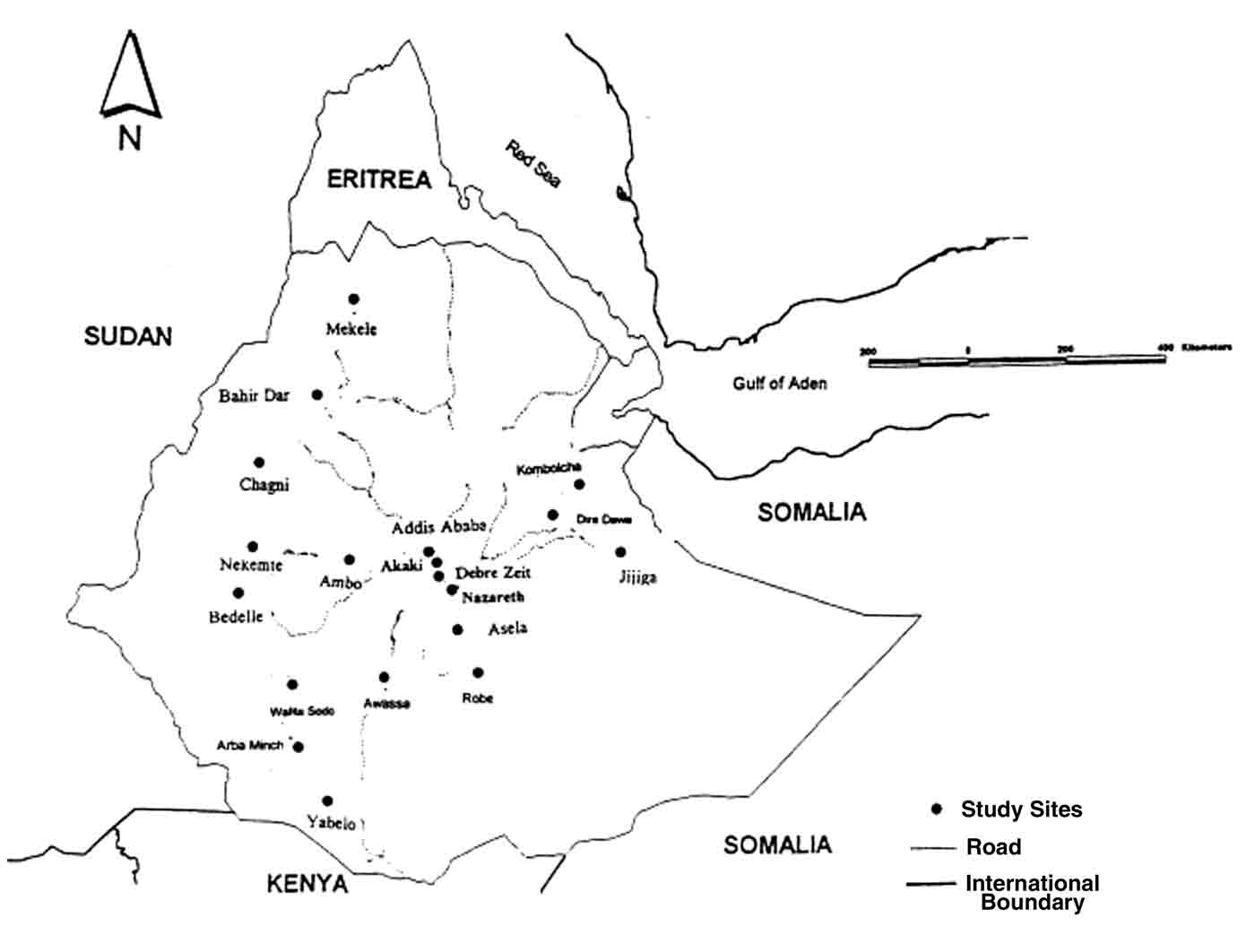
The study sites, including those for the incidence study, were indicated by the map of the country. The period prevalence and the incidence of EL in both towns were indicated by percentages.
RESULT
The retrospective analysis of the data revealed that EL and UL occur in most parts of Ethiopia (Fig. 1). Both types of lymphangitis in horses were reported from all 16 regional veterinary clinics, except that EL was not reported in horses from Yabelo or UL from Arba Minch. However, in mules, EL and UL cases were only reported from 7 and 3 regional veterinary clinics, respectively (Table 1). No cases of lymphangitis in donkeys were reported from any of the regional veterinary clinics.
The period prevalence (number of cases at the beginning plus the number of new cases) of EL at Debre Zeit (1850 m a.s.l.) and Akaki (2120 m a.s.l.) was 10.1%, whereas that of UL was only 4.4%. The percentage incidences of EL in Debre Zeit and Akaki were 2.2% and 1.0%, respectively (Table 2).
Morphology and Staining Characteristics
Double-contoured and oval-shaped organisms were detected, the cytoplasm of which was violet, pink, or blue, using Grams, PAS or Giemsa stains, respectively. The cytoplasm of some cells did not take any of the stains. Some cells were seen to be budding; others had a distorted cytoplasm, whereas still others had a single intact cytoplasm. Many budding, double-contoured, oval-shaped, yeast-like organisms were also seen in slides prepared from sections of active nodular tissue, stained by the hematoxylineosin (HE) or PAS techniques. The cytoplasm stained pink in PAS-stained preparations, whereas in HE-stained preparations it stained faint blue. Many types of yeast were seen concentrated in macrophages. Primary isolation was made possible in PPLO broth. These isolates hydrolyzed urea and thus confirmed to be H. farciminosum.
Growth of small, translucent, and ß-hemolytic colonies were detected on sheep blood agar. On subculturing on nutrient agar, the colonies were enlarged, cream in color, became opaque, dry, and crumbly. Grams staining of materials from these colonies revealed Gram-positive, small pleomorphic rods. The snapping nature when Corynebacterium cells divide caused formation of Chinese letters-like grouping under the microscope. The results of the various biochemical tests on the isolates are indicated in Table 3.
DISCUSSION
The preliminary longitudinal study carried out over 6 months at Debre Zeit and Akaki suggested monthly variations in the occurrence of EL. This can be explained by variations in humidity and temperature that affects the breeding of vector flies and the survival and multiplication of the mycelial form of H. farciminosum.10 According to Gabal and colleagues,11 EL occurs in the late summer and early winter in most countries in which it is endemic. The long incubation period and long-lasting nature of EL caused a great number of cases in January in Iraq.11 A similar finding was reported by Al-Ani and Al-Delaimi12 in Iraq. The data on UL collected from the 2 towns over the 6 months was not sufficient to allow a comparison between the 2 towns.
Both EL and UL occurred widely in Ethiopia. The fact that cases of lymphangitis were recorded in horses from almost all the regional veterinary clinics but in mules from only 3 clinics suggested that horses are more susceptible to the disease than mules. Similar results have been reported by Fraser and Maays13 and Sewell and Brocklesby.14 Chafing the legs together, uneven shoeing, and improper harnessing could be predisposing factors to lymphangitis in horses, particularly in carthorses.
The observed microscopic and cultural characteristics of both the yeast and the mycelial forms of H. farciminosum were consistent with the descriptions by Awad,15 Gabal and colleagues,16 Soliman and colleagues,17 Euzeby,9 and Guerin and colleagues.5 Primary isolation was difficult because of contamination with saprophytic fungi and the slow-growing nature of the organism. However, it was successfully achieved in PPLO broth and Sabourauds dextrose agar enriched with 10% horse serum. Hydrolysis of urea by the fungal isolate as observed in the study, was also reported by Euzeby.9
A more detailed epidemiologic investigation into the 2 diseases in general and EL in particular is indicated so that an effective control strategy can be designed.
ACKNOWLEDGMENTS
The authors thank
Dr. Fiseha Tareke and Professor Fiseha Gebreab for their technical advice.
Their sincere appreciation also goes to senior staffs of the Department
of Microbiology, Faculty of Veterinary Medicine, Addis Ababa University,
for their cooperation, provision of facilities, and lab work.
REFERENCES
1. National Animal Health Research Program Strategy Document. Addis Ababa: Ethiopian Agricultural Research Organization (EARO); 1999:146.
2. Fielding D, Pearson RA: Donkeys, mules and horses in tropical agricultural development. Proceedings of a Colloquium organized by the Edinburgh School of Agriculture and the Center for Tropical Veterinary Medicine of the University of Edinburgh, Scotland; September 36, 1991.
3. Radiostits OM, Blood DC, Gay CC: Epizootic Lymphangitis and Ulcerative Lymphangitis in Veterinary Medicine: A Textbook of the Disease of Cattle, Sheep Pig, Goat and Horses, 8th ed. Bailliere Tindall; 1994:655656, 11671169.
4. Solomon H: Animal Health Review in Ethiopia (19721979). Addis Ababa: Department of Veterinary Service Division, Ministry of Agriculture; 1980.
5. Guerin C, Abebe S, Touati F: Lymphangite epizootique du cheval en Ethiopie. Journal of Mycology 2:15, 1992.
6. Addis Ababa: National Metrology Service Agency (NMSA); 2000.
7. Carter GR: Diagnostic Procedures in Veterinary Bacteriology and Mycology, 4th ed. Charles Thomas; 1984:318328.
8. Diagnostic Procedures for Epizootic Lymphangitis. Rome: Organization Internationale des Epizootes (OIE); 1996:457459.
9. Euzeby I: Mycologe medicale compare tome I, 1st ed. Collection Foundation Marcel Herieux; 1992:432439.
10. Gabal MA, Hennager S: Study on the survival of Histoplasma farciminosum in the environment. Mykosen 26:481487, 1983.
11. Gabal MA, Hassan FK, Siad AA, Kaarim KA: Study of equine histoplasma farciminosii and characteristics of Histoplasma farciminosum. Sabouraudia 21:121127, 1983a.
12. Al-Ani KF, Al-Delaimi KA: Epizootic lymphangitis in horse: clinical, epidemiological and hematological studies. Pakistan Veterinary Journal 6:96100, 1986.
13. Fraser MC, Maays A: Epizootic lymphangitis, ulcerative lymphangitis. In: Merck Veterinary Manual, 6th ed. Rahway, NJ; 1986:56, 466.
14. Sewell MH, Brocklesby DW: Equine Histoplasmosis in the Handbook on Animal Diseases in the Tropics, 4th ed. Bailliere Tindall; 1990:364367.
15. Awad FI: Studies on epizootic lymphangitis in the Sudan. J Comp Pathol 70:457463, 1960.
16. Gabal MA, Hassan FK, Al-Siad AA, Karim KA: Study on equine histoplasmosis epizootic lymphangitis. Mykosen 26:145151, 1983b.
17. Soliman R, Saad MA, Refai M: Studies on histoplasma farciminosii in Egypt. Mykosen 28:457461, 1985.
Figure 1. Epizootic lymphangitis and ulcerative
lymphangitis occur in most parts of Ethiopia.
Table
1. Frequency
of Occurrence of Lymphangitis in Horses and Mules in 17 Regional Veterinary
Clinics
*Case(s)
detected/
Clinic Altitude no. of visits UL/EL
Addis Ababa
2408 3/11 3/11
Ambo 2000 4/5* 3/5
Arba Minch 1500
0/7 3/7
Asela 2000 5/11 7/11
Awassa 1000 2/3 2/3
Bahir Dar 1790 7/11 8/11
Bedelle 2000 4/8 4/8
Chagni 1000 1/4 ?
Dire Dewa 1160
5/11 7/11
Jijiga 1000 3/5 4/5
Kombolcha 1903
4/11 5/11
Mekele 2200 1/4 2/4
Nazareth 1000 3/8 6/8
Nekemt 2000 3/9 2/9
Robe 2300 4/7 6/7
Walita Sodo
2000 6/9 4/9
Yabelo 2000 2/7 1/7
Total
* Numerator: Number of years positive(s)
were diagnosed; denominator: total number of years each clinic was visited.
Included
cases in mules .
Table 2. A Prevalence Study in October and a 5-Month Incidence of Epizootic Lymphangitis in Debre Zeit and Akaki
Debre Zeit Akaki
No. positive/ No. of No. positive/ No. of
Months no. examined deaths no. examined
deaths
October 1995* 28/600 2 5/500 1
November 1995 15/572 3 4/495 0
December 1995 13/557 2 3/491 1
January 1996
18/544 2 6/488 0
February 1996
6/526 1 6/482 0
March
1996
8/520 1 3/476 0
Percentage incidence 2.2% 1.0%
*No. positive/no. negative.
Prevalence.
Table 3. Biochemical Test Results of the 4 isolates of Corynebacterium pseudotuberculosis
Isolates Biochemical test results
Hemolysis Catalase Glucose Maltose Trehalose Nitrate Arginine Starch Urea Growth on
on blood agar test ferm. Ferm. Ferm reduction test hydro hydro. hydro. tellurite agar
151D - + + + - - + + + +
061D - + + - - - + + + +
120D - + + + - + + - - +
154A - + + + - - + + + +
+, positive; -, negative; hydro, hydrolysis; ferm, fermentation.
ISSN# 1542-2666